SEPTARIA OF ULYANOVSK REGION AS JEWELRY AND CRAFTS MATERIAL
D.A. Petrochenkov
RGGRU, Moscow.
Together with ammonites, marl nodules contain cores of calcite of various colors, powers and textures (Fig. 1). Such nodules are known as septaria. Often septaries are made only with calcite veins and do not contain fossil shells.
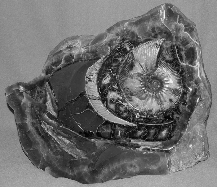
Fig. 1. Septarium containing ammonite (Goteriv tier). 35х25sm.
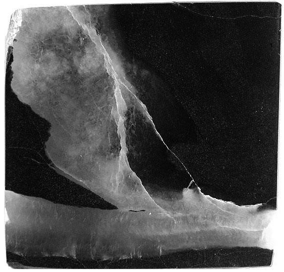
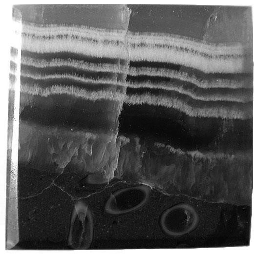
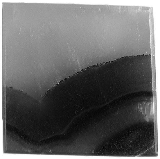
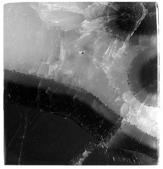
Fig. 2. Characteristic types of textures of calcite veins of septaries. 2x2sm.
It is suggested that nodules with shells formed where the latter were buried along with the soft body of the mollusk. "Empty" nodules arose around soft bodies that fell from shells. As a result of the diagenesis, the nodules were broken inside with drying cracks (compactions) and filled with mineral substance. The main volume of septaries is associated with Gotherian deposits of the lower Cretaceous.
Septaries are used to make countertops, balls, caskets, watch stands, as well as jewelry. Inserts from septaries and ammonites are externally close and received the common trademark simbirzite. Nevertheless, septaries have a number of mineral and structural features.
According to radiographic analysis, the carbonate veins of septaries consist of calcite, pyrite is noted in individual samples, and traces of organic matter and dolomite are also present. The containing rock is a marl.
Septaries have a rounded or oval shape. Their size can vary from a few centimeters to 1-2 m. The width of calcite veins in septaries varies from several mm. up to 10 or more sm. The pattern of veins is very diverse and has high decoration. The veins radially diverging from the center of the septum are characteristic, wedging out at the surface, which intersect with a series of concentric veins and veins. Sometimes the outer part of the septum sphere is framed by calcite residential. A characteristic feature of calcite veins is their symmetrical structure and clear, uneven contacts with the marl. In the central part of large veins, jeodes are often formed.
Despite the relatively simple mineral composition, carbonate cores have a wide range of color shades. The most common combinations are orange, yellowish-orange, yellow, reddish-orange colors, of varying saturation. Sometimes greenish shades are observed. Color zone transitions can be either gradual or contrasting. Zones with such colors are translucent, which creates a volumetric perception of the stone pattern.
The next most common color is brown, different shades and saturation. Zones of this color, as a rule, are located in the salbands of the strands and have clear borders with light-colored calcite. Areas of brown color are less transparent. Dark-colored areas are opaque or translucent in thin plates.
Opaque white, yellowish-white zones 1-2 to 5 mm wide are characteristic of calcite strands. They can occupy both central and peripheral parts of the cores. In the latter case, they usually follow brown-colored zones or form a sequential alternation with them.
The marl has a uniform dark gray to black color and is completely opaque.
Small crystals of pyrite are often observed in calcite veins. Sometimes they form thin (0.1-0.5 mm, rarely up to 3 mm) veins, usually located on the border of dark and light-colored zones. Pyrite has an isometric shape in the form of cubes, octahedra, pentagon-dodecahedra. The size of the crystals is usually tenths of a mm and in rare cases reaches 1-2 mm.
Note that ammonites are characterized by the constant presence of pyrite veins. Often pyrite completely replaces ammonite shell. Another external distinguishing feature of septaries is the absence of aragonite veins of the partitions and walls of the shell.
For calcite veins, septaries are primarily characterized by a striped texture formed by alternating different-colored layers (Figure 2). The width of the layers of yellow and orange colors can reach several cm, dark brown and white does not exceed the first mm. The number of alternating layers from one edge of the core is at least three, the maximum can reach 10 or more. Other types of textures are observed in certain areas of the veins. Of these, massive and spherolite can be distinguished (Figure 2).
The calcite layers are formed by prismatic, elongately prismatic, pole crystals with a fibrolite structure (Figure 3). In some areas, isometric forms of crystals with a granoblast structure are observed. The calcite layers can be separated by chains of fine pyrite crystals. In other cases, the contact of the layers is made with finely dispersed calcite.
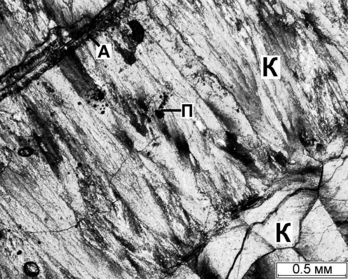
Figure. 3. Calcite streaks септариев in the shlifakh. Are crossed by Nicoli. Calcite (K), pyrite (P), aragonite (A)
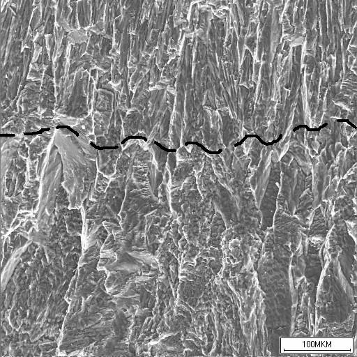
Figure. 4. Contact of yellowish-white (upper part of the picture) and brown (lower part) calcite.
Calcite crystals form sheath-shaped, radial-radiant and parallel aggregates, as well as polysynthetic doubles. In crossed nicks, they are characterized by undulating fading. The size of calcite crystals varies, as a rule, from 0.2-0.5 mm to 1-1.5 mm along a long axis, in rare cases it reaches 3 mm (Fig. 3). Prismatic calcite crystals fused to each other along the C axis reach a size of 7-8 mm. The cross-sections of the crystals are tenths and hundredths of a mm. In the isometric form of crystals, their size varies, usually in the range of 0.1-0.5 mm. Calcite size tends to increase towards the center of the core.
Pyrite occurs in the form of isometric crystals with a size of 0.1-0.5 mm. Sometimes pyrite can be observed in the form of scattered microcrystalline (0.01-0.03 mm) inclusiveness (Fig. 3). In addition to pyrite, there is a rare intricacy of organic matter. Small fragments of the pearlescent layer of shells trapped in the solution were also observed.
According to spectral semi-quantitative analysis, calcite veins in septaries contain an insignificant number of elements determined above the sensitivity of the method. In addition to the basic Mg and Ca, Si, Al, P, Na, Sr are contained in small amounts. Slightly larger values (up to 1%) are characteristic of Mn and Fe.
The results of local X-ray spectral analysis showed fairly stable contents in the calcite veins of septaries Mg, P, Ca, Mn and O in both different samples and differently colored zones. Significant content fluctuations are observed for Fe. It can be noted that zones of brown color of various shades contain Fe much less than yellow and orange. Maximum Fe values are contained in white opaque zones. These trends, but less pronounced, are noted for Mn. In general, the distribution of elements in the calcites of septaries and ammonites is close, which indicates the chemical homogeneity of the solutions.
Calcite septum veins with alternation of brown translucent and yellowish-white opaque layers were examined on a Tesla BS-301 raster electron microscope. At an increase of 250, the brown translucent layer is composed of highly elongated prism crystals of calcite (Figure 4). Their size along the axis length is 0.5-0.8 mm, with a width of 0.1-0.05 mm.
The picture shows the complex branching structure of the crystals themselves. Cleavage is well expressed on the chips. Small impurities Mn and Fe are recorded on the energy spectrum in calcite, and in individual measurements, on the sensitivity face of the P concentration method.
The opaque layer of yellowish-white color is composed of small tablate crystals of subparallel calcite. Radial radiant microstructure is traced. Crystal size already varies within 0.1-0.05 mm along axis length and 0.02-0.008 mm across. On contact with the brown layer, the crystal size may increase (Figure 4). With an increase of 800, the tabular shape of the crystals is clearly manifested, the presence of individual small pores on their surface and elongated micro-voids between the crystals. There are slightly higher Fe and Mn concentrations in this layer than in the previous one.
Thus, the outwardly different layers of calcite in the septaries differ primarily in the size and structural features of the crystals. A certain effect on the color of the layers may have Fe and Mn contents. Note the uniform mineral composition with these increases.
Examination of samples in a Tesla BS-500 transmission microscope with high resolution showed that the main matrix is built of different-sized blocks densely laid in relation to each other. At the same time, their crystallographic orientation does not coincide as a whole. This structure is characteristic in the recrystallization of a gel with numerous embryos disoriented in space.
From microimplements, plate-like gold secretions (3x8 and 2x3 microns) were found, located at the border of different size blocks, graphite particles up to 4 microns. In areas with a fine-block structure of the matrix, layered aluminosilicates up to 20 μm in size are quite often present. The listed secretions are characterized by sections of blocks that were not exposed to secondary effects. Therefore, it can be assumed that the particles of gold, graphite and laminated aluminosilicates are initially entrained in the gel.
In areas with loose laying of blocks, with the development of pores, cracks, bacteria replaced by FeO are observed. Leaching of the surface layer occurred here, which could lead to the appearance of new inclusions microphases. Thus, on the rough surface of the matrix, the extraction of vernadite with a size of 3x4 microns and magnetite particles with uneven edges of up to 6 microns is observed.
Thus, septarian calcite is characterized by a homogeneous structure and a small content of micro-impurities phases, which significantly distinguishes it from calcite in ammonites.
